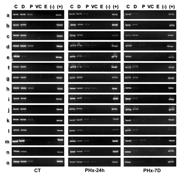
Figure 4.
Positional mapping relative to the NM of specific target sequences. The position relative to the NM of specific target sequences along the 162 kbp rat albumin gene-family genomic region was determined by PCR. Nucleoids from rat hepatocytes were treated with DNase I (0.5 U/ml) for different times (Figure 2). The residual NM-bound DNA in the partially digested samples was directly used as template for PCR amplification of the chosen target sequences (a - o). The specific amplicons were resolved in 2% agarose gels and stained with ethidium bromide (0.5 μg/ml). C, 0' digestion-time control. The amplicons were scored either as positive or negative as a function of endonuclease digestion time and for each topological zone relative to the NM, depending on whether or not they were detected by a digital image-analysis system (Kodak 1D Image Analysis Software 3.5) using the default settings. Topological zones relative to the NM: D, distal; P, proximal; VC, very close; E, embedded within the NM. (-) Negative control (no template); (+) positive control (pure genomic DNA as template). CT, control G0 hepatocytes; PHx-24 h, hepatocytes 24 h after partial hepatectomy; PHx-7 D, hepatocytes 7 days after partial hepatectomy. The amplification patterns were consistently reproduced in separate experiments with samples from independent animals (n ≥ 3).

