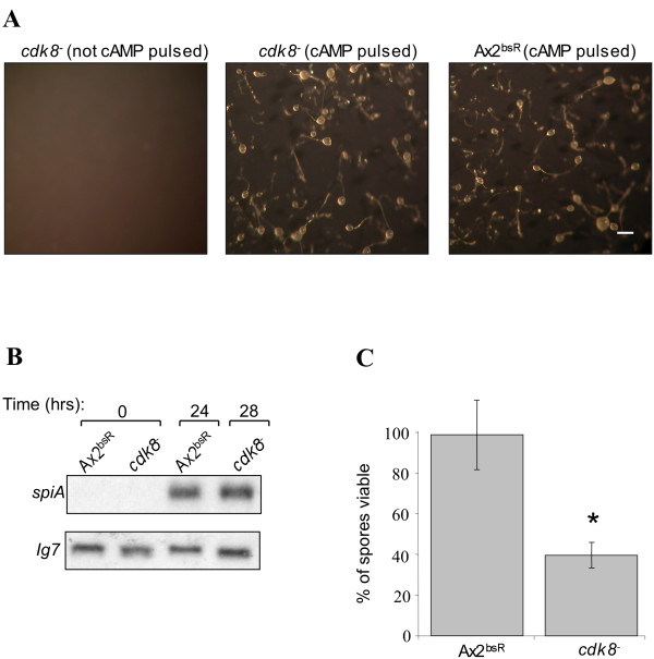
Figure 2.
Late developmental phenotype of cdk8-2 cells. (A) Cells were developed in KK2 buffer at 1 x 10⁷cells/ml with or without cAMP pulsing (50 nm cAMP added to the suspension every 5 minutes). Each strain was shaken for 6hrs at 150 rpm at 22°C before being spread onto filters at a density of 3 x 10⁶ cell/cm2. Photographs were taken after 28hrs. The scale bar in the right hand panel represents 200 μm. (B) Expression of SpiA in cdk8-2 cells. Fruiting bodies formed after cAMP pulsing were harvested after 24hrs or 28 hrs. RNA was extracted from these samples and resolved on a 1% formaldehyde gel, transferred to a nylon membrane and probed with a 32P labelled fragment of the spiA gene. The blot was reprobed with a 32P labelled fragment of the IG7 gene so as to control for loading. (C) Viability of cdk8-2 spores. Fruiting bodies were formed by developing cells in shaken suspension with cAMP pulsing prior to plating. Equal numbers of spores from each strain were treated with heat and detergent and spread onto a bacterial lawn. Colonies resulting from hatching of viable spores were counted after 4-5 days. Each bar represents the mean (±standard deviation) of three independent experiments. Results showing statistically significant difference from Ax2bsR are marked with a * (p < 0.05 by student t-test).

