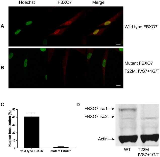
Figure 5. Expression of endogenous FBXO7 in human fibroblasts.
(A, B) For immunofluorescence, the FBXO7 protein is visualized in red by using a mouse primary anti-FBXO7 antibody and a Cy3-coupled secondary anti-mouse antibody. The nucleus (Hoechst staining) is depicted in green. (scale bars, 10 µm). (A) normal control; (B) Dutch PARK15 patient with T22M and IVS7+1G/T mutations. (C) quantification of percentages of cells showing mainly nuclear localization of FBXO7. (D) Western blotting analysis of fibroblasts from a normal control and the Dutch PARK15 patient.

