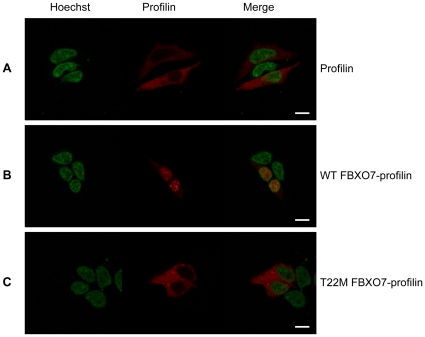
Figure 6. The N-terminus of FBXO7 confers nuclear localization to profilin.
In panel (A), Profilin, a well-known cytosolic protein, is visualized by the red mCherry signal; the nucleus is depicted in green (Hoechst staining). Expressing the first 40 amino acids of WT-FBXO7 isoform 1 in front of mCherry-labeled profilin changes the localization of profilin from the cytoplasm to the nucleus (B). The same FBXO7 N-terminal peptide carrying the T22M-mutation found in PARK15 patients is totally devoid of this capacity (C). (scale bars, 10 µm).

