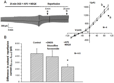
Figure 5. OGD induced VRAC activation requires Na- +loading through glutamate receptors.
A. In the presence of 40 µM AP-5 and 30 µM NBQX in the OGD solution to inhibit ionotropic glutamate receptors, the 25 min OGD induced neuronal electrophysiological changes seen in the Fig. 3A were largely attenuated. Also, the activation of VRAC in the reperfusion stage was significantly inhibited. B. Shows the differences in the outward current amplitudes between 6 min and 20 min post-OGD under the following conditions: 1) control; 2) in the presence of DNDS+ bicuculline +furosemide in the OGD and 3) in the presence of AP-5+NBQX in the OGD solution. * Indicates that the difference between the control and AP-5+NBQX groups was statistically significant at p<0.05.

