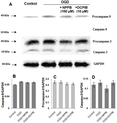
Figure 7. Short-term OGD did not change the levels of caspase-9 and -3.
A. Western blot analysis was used to detect caspase-9, caspase-3 and their precursors from lysates of slices pretreated with different conditions as indicated. 100 µg whole cell proteins in each lane were used in western analysis using anti- caspase-9, caspase-3 and GAPDH. B-D. Bar graphs show the expression levels of procaspase-9, procaspase-3 and caspase-3 that are normalized to GAPDH (n = 3). The difference was not statistically significant among different treatment groups in each category shown in B–D.

