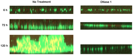
Figure 5. Susceptibility of flow cell biofilms to DNase I.
Representative z-reconstructions of RB50 biofilms grown under flow conditions for 6, 72, or 120h and imaged using CLSM for live GFP expressing cells (green) and eDNA stained with DDAO (red or yellow with co-localization). The image of untreated biofilms (left panels) were taken immediately prior to incubation with DNase I and the images of same biofilms treated with DNase I for 1.5h (left panels). Images shown here are representative of two independent experiments.

