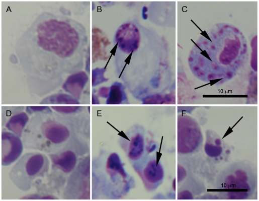
Figure 6. Histological changes related to the apoptotic process observed in hemocytes treated with UV light.
A and D: Control granulocytes and small hyalinocytes not treated with UV-light. B: Slight chromatin condensation in granulocytes observed after 3 h pt. C and F: Appearance of intracellular bodies, positive stained for DNA, inside the cytoplasm of granulocytes and hyalinocytes, respectively after 24 h pt. E: Chromatin condensation in small hyalinocytes after 6 h pt.

