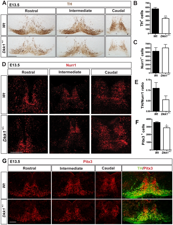
Figure 3. Dopaminergic neuron differentiation is disrupted in Dkk1 mutants.
(A) Immunohistochemistry for TH on midbrain serial coronal sections at E13.5. Scale bar = 100 µm. Quantification in (B) revealed a 40% decrease in the numbers of dopaminergic neurons in Dkk1+/− embryos when compared to Wt littermate controls (mean ± s.e.m- Wt : 568.3±31.7, N = 3; Dkk1+/− : 346.5±27.2, N = 8 p = 0.0015 ** unpaired t-test). (C, D) The Nurr1+ dopaminergic precursors are not affected in Dkk1+/− embryos (mean ± s.e.m- Wt : 5233±857.0 N = 2; Dkk1+/− : 6019±840.8 N = 3). Scale bar = 100 µm. (E) The TH/Nurr1 ratio indicated a differentiation deficit in the Dkk1+/− embryos (mean ± s.e.m- Wt : 0.109±0.027, N = 2; Dkk1+/− : 0.055±0.018, N = 3). (F, G) Expression of Pitx3 was reduced in Dkk1+/− embryos compared with wild-type littermates (mean ± s.e.m- Wt : 3574±99.02, N = 3; Dkk1+/− : 2874±216.4, N = 3, unpaired t-test p = 0.042*). Scale bar = 100 µm.

