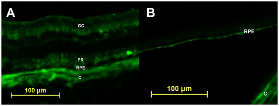
Figure 3. Fluorsecent light micrographs of retinal cross sections.
A. Treated eye of a Dutch Belted rabbit demonstrating immunofluorescence likely occuring at the level of choroid (C), retinal pigmented epithelium (RPE), photoreceptors (PR), and retinal ganglion cells (GC). B. Untreated eye of a Dutch Belted rabbit with autofluorescence seen at the level of choroid (C) and retinal pigmented epithelium (RPE).

