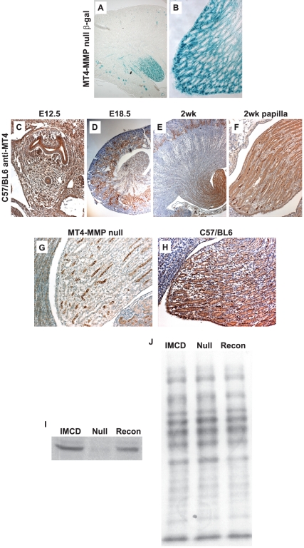
Figure 1. MT4-MMP localization in the mouse kidney.
(A–B) β-galactosidase staining kidneys from 5week old MT4-MMP null mice shows staining in the cortex and medulla (A, 50X DIC) with the most intense staining seen in the papilla (B, 200X DIC). (C–F) Staining with a specific antibody directed against MT4-MMP in wildtype mice shows expression throughout the metanephric mesenchyme and ureteric bud at E12.5 (C, 100X). MT4-MMP expression is abundant in the cortical regions during embryonic development (E18.5, D, 50X), but by 2weeks of age, most of the expression is localized to the papilla regions (E–F, 50X and 200X, respectively). (G–H) Staining with a specific antibody directed against MT4-MMP in MT4-MMP null and wildtype mice, demonstrating specificity of the antibody for MT4-MMP (200X). (I–J) Immunoblot of inner medullary collecting duct cells derived from wild type mice (IMCD), MT4-MMP null mice (Null) or IMCD cells from MT4-MMP null mice reconstituted with MT4-MMP (Recon), illustrating specificity of the antibody for MT4-MMP (I). Ponceau staining was performed on the nitrocellulose of the immunoblot to demonstrate equal loading of the lysates (J).

