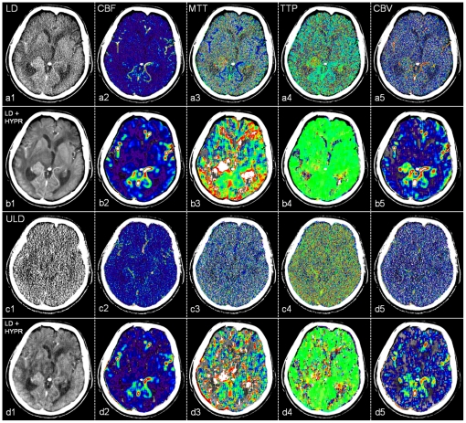
Figure 3. Low dose (LD, a), HYPR-LR-post-processed low dose (LD+HYPR, b), ultra low dose (ULD, c) and HYPR-LR-post-processed ultra low dose (ULD+HYPR, d) brain perfusion CT of a 75-year old male patient with a lung cancer metastasis adjacent to the right thalamus and chronic left frontal infarction.
Last image of the 60 s time series (1) and the normalized cerebral blood flow (CBF, 2), mean transit time (MTT, 3), time to peak (TTP, 4), cerebral blood volume (CBV, 5) maps. The utilized software does not use reduced matrix reconstructions or spatial smoothing, the images are left noisy. The pathology is recognizable in a, b and d with excellent subjective image quality and low noise in b. No diagnosis possible in c.

