Fig. 1.
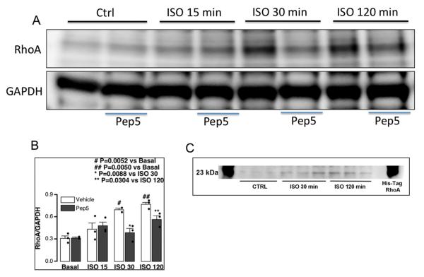
Isoflurane exposure increases RhoA activation in DIV5 neurons in vitro. Primary neurons (4-7 days in vitro – DIV4-7) were exposed to 1.4% isoflurane for 15, 30, and 120 min with and without pretreatment with TAT-Pep5. Immunoblot analysis (A) demonstrated that RhoA was enhanced at both 30 min and 120 min following isoflurane exposure (n = 3; # p = 0.0052 vs. basal, ## p = 0.005 vs. basal) compared to control (Ctrl). Pretreatment with TAT-Pep5 (15 min; 10 μM) significantly decreased isoflurane-mediated RhoA activation at both 30 min and 120 min (n = 3; * p = 0.0088 vs. isoflurane 30, ** p = 0.0304 vs. isoflurane 120). Quantitation of the data is represented in the figure panel (B). An immunoblot showing that the RhoA band (~23-25 kDa) corresponds to the His-Tag RhoA loaded control (C). RhoA values were normalized to glyceraldehyde 3-phosphate dehydrogenase. Error bars, standard error of the mean (s.e.m.).

