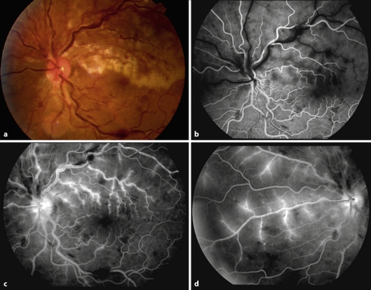
fig. 1.
Fluorescein angiogram 24.0 seg, 3:53 and 8:32. Fluorescein angiography shows delayed arterial and venous filling, large areas of ischemia in the superior quadrants, retinal hemorrhages, periphlebitis, macular edema, upper trunk venous occlusion and lower trunk venous stasis associated with occlusion of the superior temporal arterial branch.

