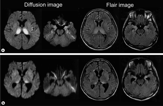
Fig. 2.
Brain MRI findings. a Images on the 42nd hospital day showed high-intensity lesions in the bilateral thalamus and bilateral medial temporal lobes. b Images on the 64th hospital day revealed improved abnormal high-intensity lesions in the bilateral thalamus but progressive atrophy of the bilateral medial temporal lobes.

