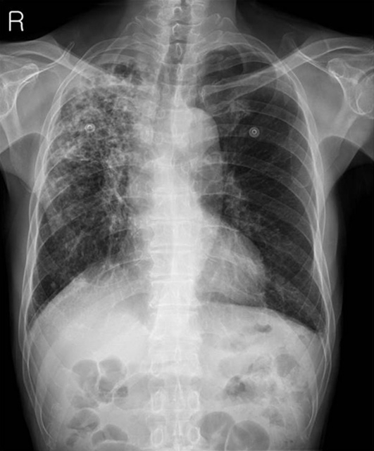
Fig. 1.
A chest radiograph taken at admission demonstrates whitish patch consolidations on the right upper lung field suggesting pulmonary tuberculosis. The right middle and lower lung fields are also involved with patch consolidations but show much less whitish patches than those of the right upper lung field. Contrasting to the right lung field, the left lung field does not show abnormal findings. There are no vascular abnormalities and no bony thorax abnormalities.

