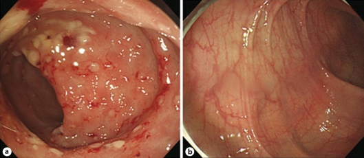
Fig. 2.
a Sigmoidoscopy demonstrates multiple whitish to yellowish plaques, 5 to 8 mm in size. Many whitish to yellowish plaques are aggregated with much exudate and some fresh blood or scattered on the whole sigmoid colon. Multiple hyperemic erosive lesions are also scattered on the whole sigmoid colon with edematous mucosa. b Sigmoidoscopy, after 2 weeks of metronidazole treatment, shows that the multiple whitish to yellowish plaques and accompanied small erosions, responsible for bleeding, have disappeared completely. Pinkish mucosa with fine normal vascular patterns, which is not observed in a due to edema, is observed.

