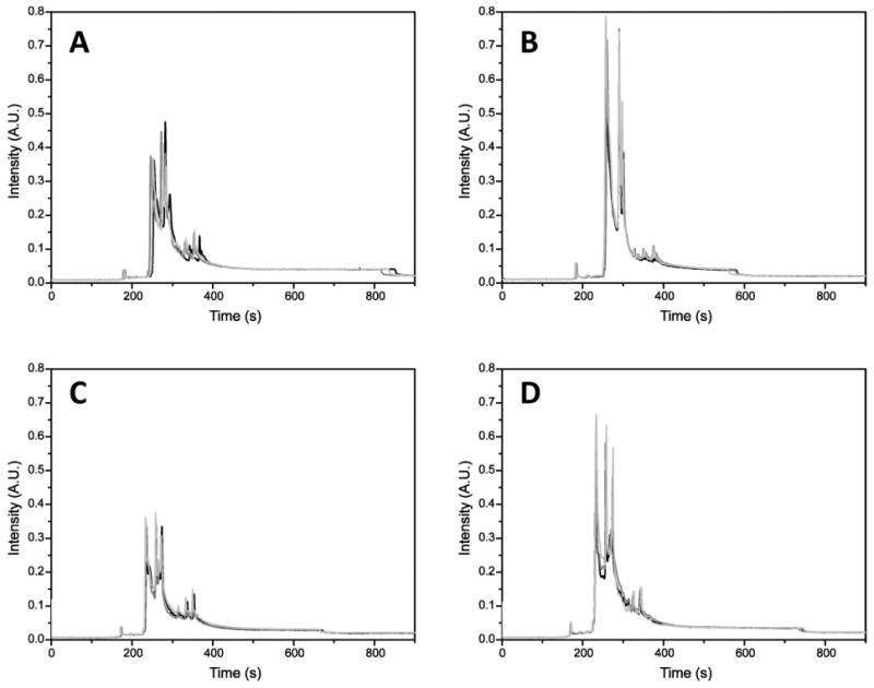
Figure 1.

Electropherograms of mitochondrial proteins labeled with Alexa 488 hydrazide. A, Young, fast-twitch. B, Old, fast-twitch. C, Young, slow-twitch. D, Old, slow-twitch. Separation, −570 V/cm; hydrodynamic injection, 11 kPa, 4 seconds; sieving matrix, 20 mM Tris, 20 mM tricine, 8% dextran (462 kD), 0.5% sodium dodecyl sulfate, pH 8. The samples were analyzed in triplicate. Capillary conditioning, fluorescence labeling, and detection are described in Materials and Methods. A.U. = arbitrary units.
