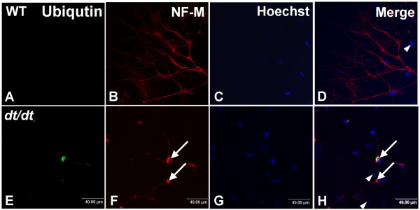
Figure 7.
Immunoreactivity of ubiquitin and NF-M in cultured neurons from wild-type and dt/dt mice. Cultured neurons were double-labeled with antibodies against ubiquitin (green) and neuronal intermediate filament protein NF-M (red), and their nuclei were stained with Hoechst 33342 (blue). The ubiquitin-positive reaction was hardly noticeable in neurons of wild-type mice (A-D). Cultured neurons with abnormal accumulations of NF-M were mostly observed in the proximal region of axons and within cell bodies of cultured sympathetic neurons from dt/dt mutants (F, arrows). Some neurons with NF-M accumulations could also be labeled with the antibody against the ubiquitin (E-H). Some smaller nuclei of non-neuronal cells were also observed in the primary culture (arrowheads, D and H). Scale bars = 40 μm.

