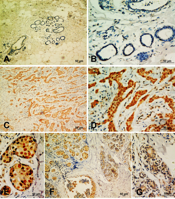
Figure 1.
Increased expression of Bmi-1 in breast cancer tissues. (A, B) Bmi-1 showed no or weak staining in the adjacent non-cancerous tissue. (C, D) Strong Bmi-1 staining was detected in the primary breast cancer tissue. (E, F, J) Cancer showed high Bmi-1 expression and nuclear staining, whereas the adjacent mammary gland lobule showed low expression and cytoplasmic staining (100X and 400X).

