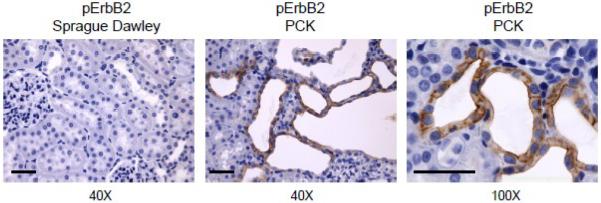
Fig. 2. pErbB2 expression in normal and cystic rat kidney tissue.
Representative immunohistochemical staining for pErbB2 detection in kidney cortical sections of Sprague Dawley (a) and PCK (b, c) rats. Original magnifications are 40× and 100× (scale bars: 50 μm). Tissue sections were stained with anti-pErbB2 (Phospho-HER2/ErbB2 (Tyr1221/1222); 1:100; Cat. #2243; Cell Signaling Technology Inc., Boston, MA).

