Figure 4.
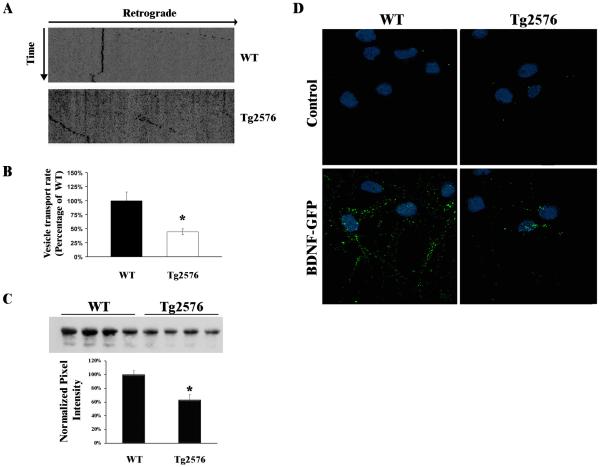
The retrograde transport of BDNF-GFP is reduced in Tg2576 neurons. (A) Representative kymograph of BDNF-GFP vesicles within axons of WT (top) and Tg2576 (bottom) neurons. The time arrow represents 5 min. and the retrograde arrow indicates 65.2 μm. (B) Individual BDNF-GFP vesicle velocities were assessed using ImageJ (NIH). The average velocity of BDNF-GFP vesicles was significantly reduced in Tg2576 neurons compared to WT (*p<0.005, n=37 for WT, n=24 for Tg2576). (C) Lysates from somal compartments were immunoprecipated with rabbit anti-GFP antibody (Invitrogen) followed by Western blot analysis with mouse anti-GFP antibodies and revealed a BDNF-GFP band that was decreased in Tg2576 somal lysates when compared to WT. Quantification of BDNF-GFP on the somal side demonstrated a significant reduction (*p=0.003, n=4) in net BDNF-GFP transport in Tg2576 vs WT neurons. (D) Immunocytochemical analysis of Tg2576 and WT somal compartments. In the absence of BDNF (Control, Top panels), BDNF-GFP-labeling was not detected in somal chambers, but after 2hr, BDNF-GFP levels were increased in the somal compartment of WT, but to a lesser extent in Tg2576 (Lower panels).

