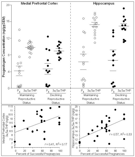
Fig. 1.
Depicts concentrations of progesterone (P4) and 3α,5α-THP in medial prefrontal cortex (top, left) and hippocampus (top, right) of middle-aged rats whose reproductive status was in decline (n=12) or was maintained (n=14). The percentage of prior pregnancies positively correlated with concentrations of 3α,5α-THP in the medial prefrontal cortex (bottom, left) and hippocampus (bottom, right) and accounted for a significant proportion of variance in these levels.

