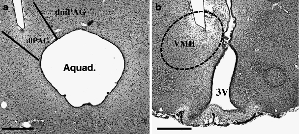
Fig. 1.

Representative low-power photomicrographs of 30-μm-thick frontal sections from the brain of a rat subjected to stereotactic implantation of a concentric bipolar electrode to stimulate the dlPAG (a, scale bar = 250 μm) and VMH (b, scale bar = 500 μm). The tips of the electrodes are situated within the respective targets. Aquad aqueduct of Sylvius, dmPAG dorsomedial periaqueductal grey, dlPAG dorsolateral periaqueductal grey, 3V third ventricle, VMH ventromedial hypothalamus
