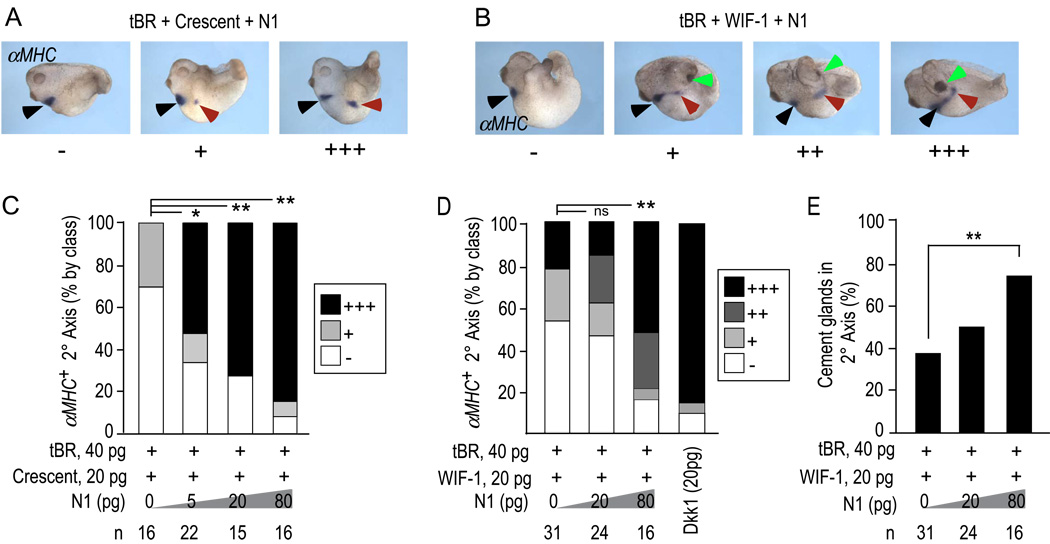
Figure 4. N1 synergizes with WIF-1 and Crescent to induce heart and cement gland.
A, B) Examples of embryos as in Fig. 3 cultured to stage 28–30 and stained by in situ hybridization for αMHC to reveal developing myocardial tissue in primary (black arrow) and secondary (red arrow) body axes. N1 in conjunction with WIF-1, but not Crescent increased the incidence of cement glands (green arrows).
C, D) Quantification of the percentage of ectopic heart field induced in response to N1 plus Crescent (C) or WIF-1 (D), and according to class (+/−, +, ++, and +++) as in panels A and B. p-values were calculated by the Chi-square test [**, <0.001; *, <0.05; ns, not significant (>0.05)]. Results showing complementarity are for 20 pg of Crescent and WIF-1; similar outcomes were observed in additional experiments using 10 and 40 pg doses of both Crescent and WIF-1.
E) Quantification of the incidence of cement gland formation in response to WIF-1, N1 and tBR. N1 did not alter the lack of cement gland induction by Crescent and tBR (not shown, but see text). ** indicates p value <0.001 as calculated by Student’s T-test.

