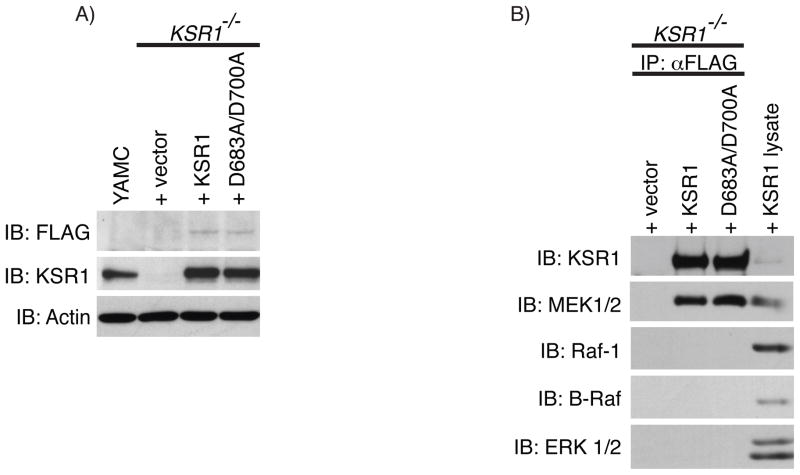
Fig. 1.
Wild-type KSR1 and kinase-inactive KSR1 associate with MEK. A) KSR1 protein expression was determined by Western blot analysis on whole cell lysates from KSR1−/− mouse colon epithelial cells expressing +vector, +KSR1, or +D683A/D700A, or from YAMC cells using the indicated antibodies. B) Co-immunoprecipitation of MEK was determined by Western blot analysis on FLAG-immunoprecipitates from +vec, +KSR1, and +D683A/D700A expressing cells that were washed in high salt immunoprecipitation buffer. Whole cell lysate from +KSR1 cells was used as a control for detecting each protein probed. Immunoblots are representatives of at least 3 independent experiments. IP, immunoprecipitation; IB, immunoblot.

