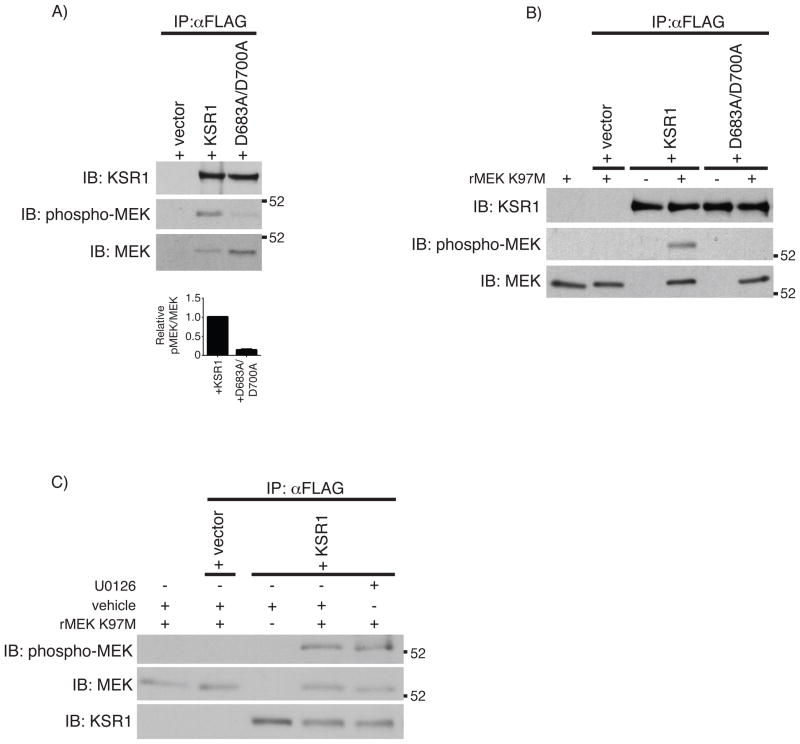
Fig. 3.
KSR1 phosphorylates MEK1 A) FLAG immunoprecipitation was performed from 1 mg protein on whole cell lysates from KSR1−/− cells expressing +vector, +KSR1, or +D683A/D700A. MEK phosphorylation was determined by Western blot analysis using the indicated antibodies. Densitometric analysis was performed on total and phosphorylated co-precipitated MEK protein and represented as the ratio of phosphorylated MEK/total MEK. Solid bars represent the mean ratio from 3 independent experiments and error bars represent the SEM. Statistical analysis was performed using a Student’s t-test comparing the pMEK/MEK ratio from +KSR1 and +D683A/D700A co-precipitations. *** P < 0.001 B) In vitro kinase assay using immunoprecipitated FLAG-tagged KSR1 proteins incubated with kinase-inactive rMEK K97M. Total protein and phosphorylated rMEK K97M were determined by Western blot analysis. C) In vitro kinase assay performed as before following a 30 minutes pre-incubation with vehicle or U0126 (10 μM). Total and phosphorylated proteins were determined by Western blot analysis. IP, immunoprecipitation; IB, immunoblot.

