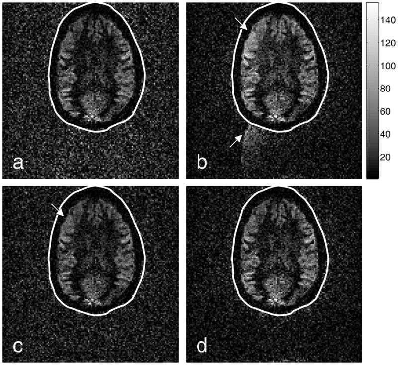
Figure 6.

Perfusion images generated from the (a) Ahn-and-Cho dynamic, (b) real-time PAGE, (c) PLACE, and (d) EPI-GESTE methods. The perfusion data is shown with a normal gray scale inside the ellipse. Outside the ellipse, the signal-void region has been amplified by a factor of four, to enhance ghost visibility. The arrows point to regions affected by Nyquist ghosts, which appear as image brightening in the upper left brain quadrant and ghost artifacts in the signal-void region.
