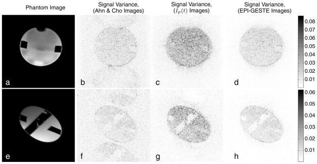
Figure 8.
Phantom (a,e) and signal variance images associated with (b,f) Ahn & Cho ghost correction, (c,g) the output of a single pMRI reconstruction channel, Ip(t), and (d,h) the final EPI-GESTE reconstruction, Ip(t) + ΨIn(t). The top row shows an axial view of the phantom. The bottom row shows a double-oblique view.

