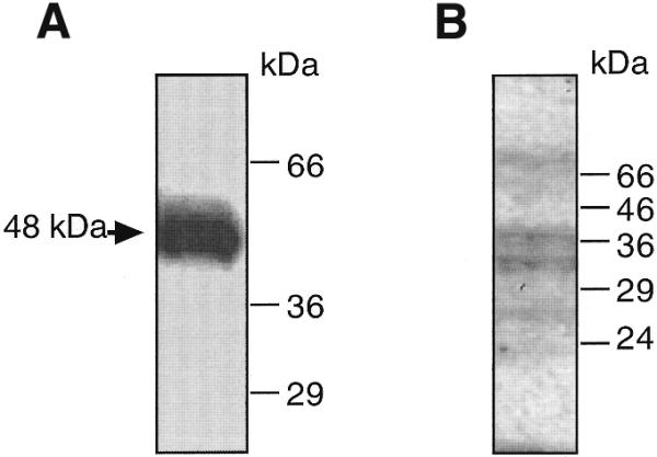
Figure 4.

An ∼48 kDa protein is UV cross-linked to the 0.17 kb rhoB promoter fragment. (A) 32P-labeled rhoB fragment was incubated with extracts from UV-irradiated cells under identical conditions to those used in the gel retardation experiments. Afterwards the reaction was irradiated with UVC light as described in Materials and Methods. Products were separated by SDS–PAGE and the cross-linked protein–DNA complex was visualized by autoradiography. (B) An aliquot of 50 µg protein from total cell extract was separated by SDS–PAGE. After blotting to nitrocellulose, southwestern analysis using 32P-labeled rhoB promoter fragment was performed as described in Materials and Methods. The autoradiogram is shown.
