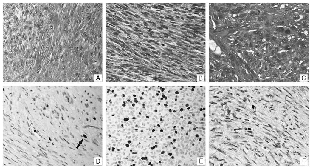
FIGURE 1.
Histologic and immunohistochemical correlates of uterine smooth muscle tumors and their pulmonary counterparts. Panel A is a representative histologic section of a leiomyoma included in this study. The microscopic features are similar to that observed in the uterus (not shown) and lung (panel B), in a woman who had a benign metastasizing leiomyoma that was noted 15 years after the removal of her uterus. Panel C, in comparison, shows the histologic features of a leiomyosarcoma that had metastasized to the lung; note the marked nuclear atypia, disorganized growth pattern, and mitotic figures. Panels D and E show representative areas of the Ki67 data for a leiomyoma (D, arrow) and leiomyosarcoma (E), respectively. The Ki67 results for the pulmonary component of the benign metastasizing leiomyomas (panel F) was equivalent to that for the leiomyomas.

