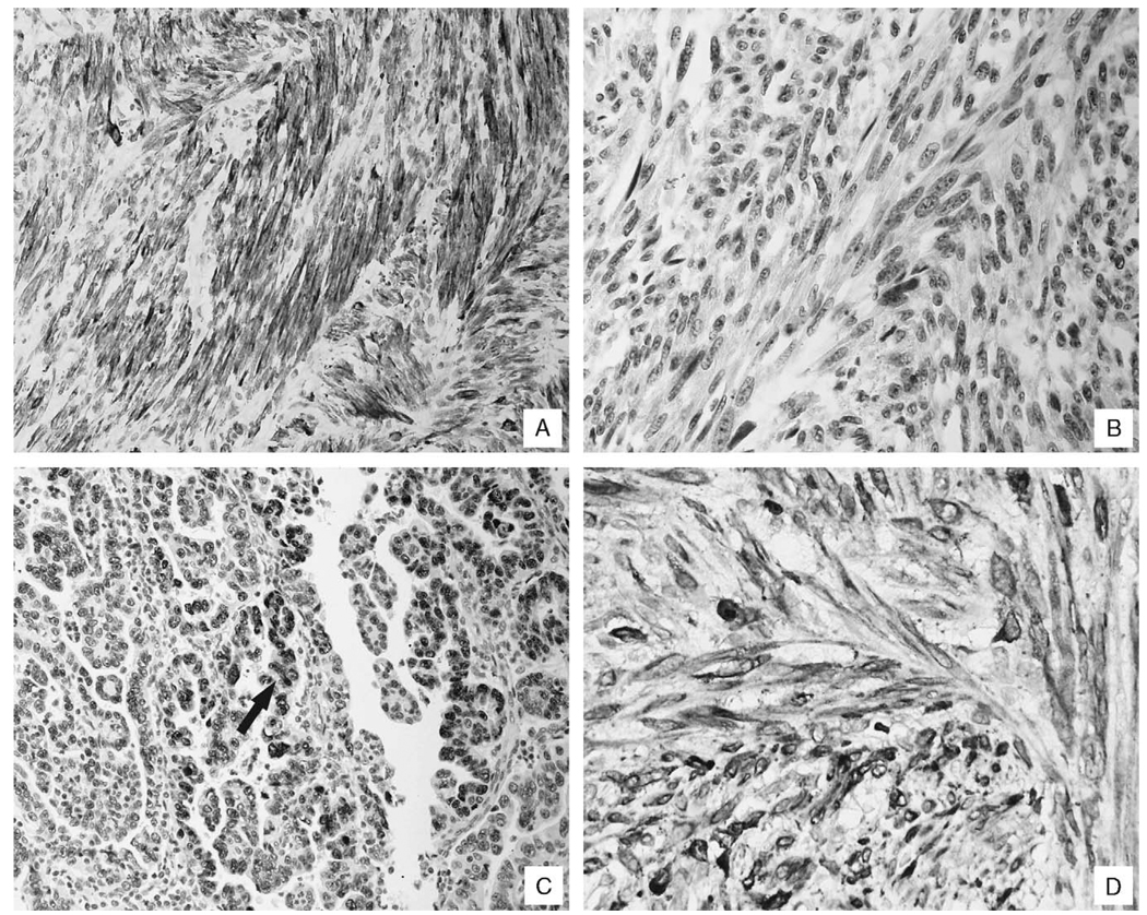
FIGURE 2.
Immunohistochemical correlates of uterine smooth muscle tumors and their pulmonary counterparts. Panel A shows a typical result for the HHF-35 immunohistochemical test for the pulmonary lesions of the benign metastasizing leiomyomas; the strong signal documents the smooth muscle origin of the lesion. Each of the benign metastasizing leiomyomas was negative for the p53 antigen (panel B). In contrast, note the nuclear-based signal, typical of most of the leiomyosarcomas (panel C, arrow). Panel D shows the strong signal for bcl-2, noted in the pulmonary lesion of the benign metastasizing leiomyoma, which is indicative of its uterine origin.

