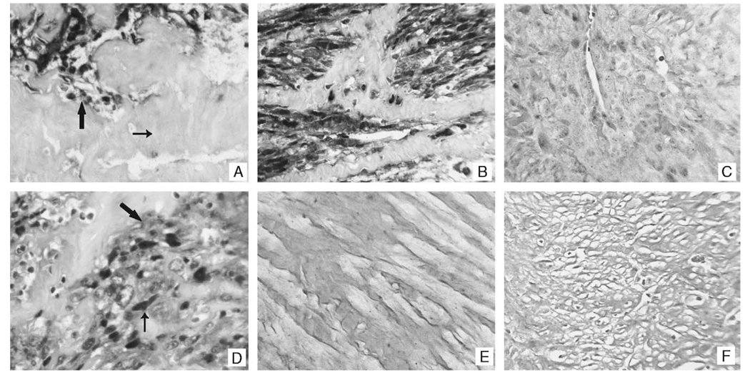
FIGURE 3.
MiR-221–expression patterns in the uterine smooth muscle tumors and their pulmonary counterparts. Panel A contains a representative example of miR-221 detection in a leiomyosarcoma of the uterus. Note the strong signal in the tumor cells (large arrow) and the lack of a signal in the adjacent fibrous tissue (small arrow). A similar pattern was evident in the lesions showing pulmonary metastasis (panel B). The signal was lost in the leiomyosarcoma if the probe was omitted (not shown) or if a scrambled lock nucleic acid probe was used (panel C). Panel D shows the miR-221 signal in the leiomyosarcomas at higher magnification; note that the signal is present in the cytoplasm (large arrow) and tends to concentrate around the nucleus (small arrow). MiR-221 was not detected by in situ hybridization in any of the leiomyomas (panel E) or benign metastasizing leiomyomas (panel F).

