
Fig. 2.
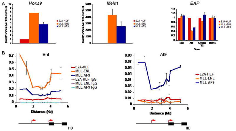
MLL fusion protein binding to target loci A) qPCR for Hoxa9, Meis1, and EAP expression in E2A-HLF, MLL-ENL, and MLL-AF9-transformed myeloblastic cell lines. Expression is normalized to GAPDH. B) ChIP-qPCR for MLL fusion partners across Hoxa9 in leukemic cell lines with or without MLL fusion proteins. Schematics of the Hoxa9 locus are depicted below the graphs. Black arrows indicate transcriptional start sites. Black boxes indicate exons.