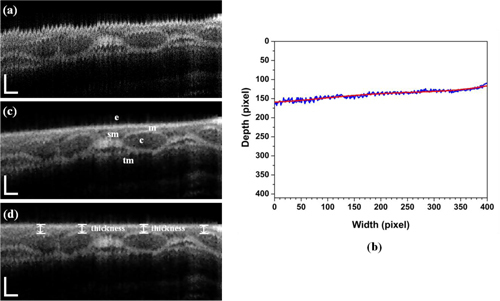
Fig. 2.

In-vivo longitudinal OCT image of the airway in a rabbit. This longitudinal images were reconstructed with 400 B-scan slices corresponding to the physical length of 8.0 mm. (a) without motion artifacts correction, (b) surface detection (blue) and result (red) after a fourth-order polynomial fit, (c) with motion artifacts correction, and (c) with flattening of the surface to measure the thickness of mucosa in normal direction from the surface; e- epithelium, m- mucosa, c- cartilage, sm- submucosa, and tm- muscularis are clearly seen. Scale bar is physically 250 μm (axial) and 500 μm (lateral).
