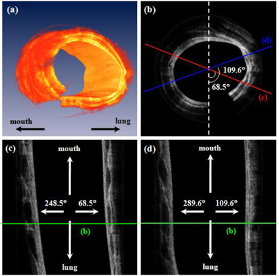
Fig. 4.

In-vivo OCT images of normal airway in a rabbit. (a) 3-D reconstructed image based on 400 B-scan slices. (b) a circumferential 2-D image (Media 1 (3.1MB, MOV) ) at one position corresponding with green lines in Fig. 4(c), and 4(d). (c) and (d) are longitudinal images, which are arbitrarily cut at 68.5° (red) and 109.6° (blue) to the vertical direction with counterclockwise (CCW) as shown in Fig. 4(b), respectively (Media 2 (3.8MB, MOV) ). Media 1 is made with moved slices from inner area to outer area. Media 2 is also a movie constructed with cut longitudinal images from 0° to 180° with respect to the vertical axis with CCW rotation.
