
Fig. 2.
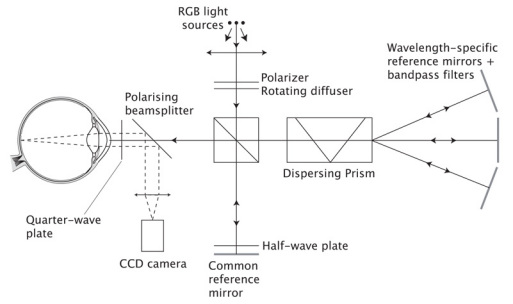
Schematic representation of the proposed Structured Illumination Ophthalmoscope using the Fizeau fringe projection technique and three wavelengths for illumination. Red, green and blue incoherent light sources are used. These are matched to the detection peaks for the detectors in the colour CCD camera and, with appropriate filtering, cross-talk is minimised. A rotating diffuser is used to enhance spatial incoherence. A beamsplitter splits the light into the two branches of a Michelson interferometer, one containing a mirror common to all wavelengths, the other containing a dispersing prism and three mirrors, one for each of the illuminating wavelengths. The fringes produced are projected onto the retina by the optics of the eye. Incoming and outgoing light from the eye is split using a polarizing beamplitter and a quarter-wave plate in front of the eye to maximise the returning light based on the eye’s birefringence. A half-wave plate in the common branch of the interferometer controls fringe modulation.