Fig. 2.
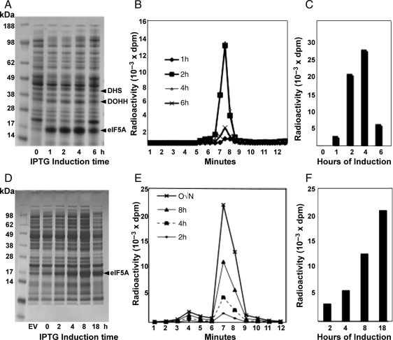
Time course of protein expression and hypusine modification upon IPTG induction at 37°C (A–C) and at 18°C (D–F). BL21(DE3)pLysS cells transformed with pST39/heIF5A/hDHS/hDOHH were induced with IPTG at 37°C or 18°C in the absence or presence of [3H]spermidine (5 μCi/ml) for the times indicated. Protein expression was examined by Coomassie-Blue staining after SDS-PAGE (A, D) and the extent of the hypusine modification was measured by measuring radioactivity in the hypusine after ion exchange chromatographic separation (B, E). Total amount of hypusine formed at each induction time point is shown as a bar graph (C, F).

