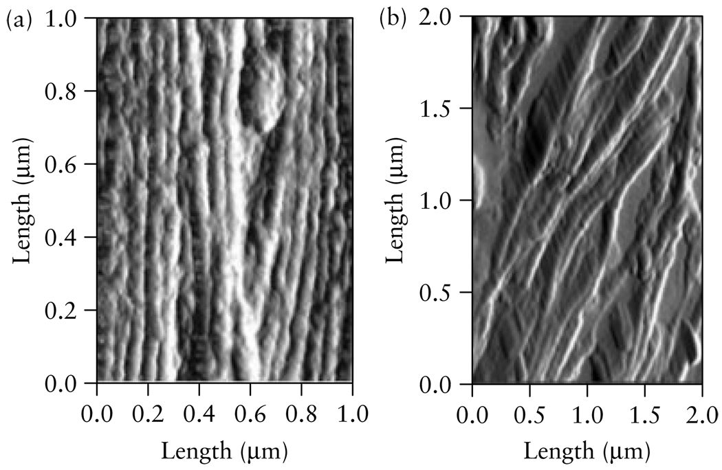
Figure 1.
Atomic force microscopy images of the cervix from (a) a non-pregnant rat, displaying highly organized, pack bundles of collagen and (b) a cervix on day 21 of pregnancy, displaying disorganized collagen with space between the fibrils. The bundles of collagen that are displayed in these images are <0.1 µm. Rats typically deliver on day 21. (Image by R. Bhargava, W. King, UIUC, and B. L. McFarlin, UIC).

