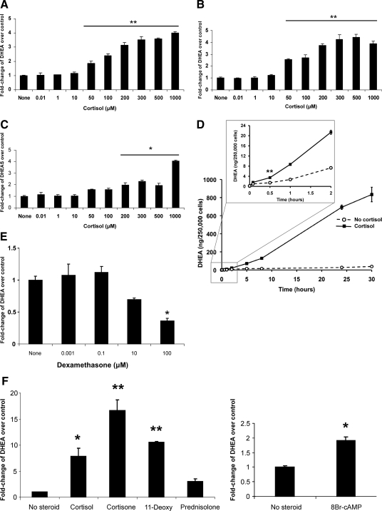
Figure 1.
Effect of cortisol upon DHEA secretion from human adrenal cells. Dose-response of DHEA secretion from NCI-H295R cells, measured by RIA (A) or by liquid chromatography tandem mass spectrometry (LC/MS-MS) (B), after exposure to cortisol for 8 h. In both panels, compared with no steroid treatment, cortisol ≥50 μm caused a significant increase in DHEA secretion. Absolute DHEA measurements, in ng/mg protein, for the no steroid treatments were 52.9 (A) and 14.3 (B), respectively. C, Dose-response of DHEAS secretion from NCI-H295R cells, measured by ELISA. D, Time course of cortisol (500 μm) stimulation of DHEA secretion, measured by RIA. The inset highlights the first 2 h of treatment, with a significant increase in DHEA at and beyond 30 min. E, DHEA secretion was not stimulated after exposure for 8 h to a wide range of dexamethasone concentrations and was significantly inhibited at the highest concentration. F, Compared with no steroid treatment, exposure to cortisone or 11-deoxycortisol (11-Deoxy) stimulated DHEA secretion, whereas exposure to prednisolone was no different than untreated cells. Treatment with 8-bromo-cAMP also showed stimulation of DHEA production. F, Cells were exposed to steroids (250 μm) or 8-bromo-cAMP (100 μm) for 24 h. DHEA measurements in E and F were done by ELISA. Each value is expressed as the mean of three replicates per group. Each experiment, except the time course (D), was performed 2–4 times with similar results. The time course (D) was repeated once, though with fewer time points, and yielded similar results. *, P < 0.005; **, P < 0.001.

