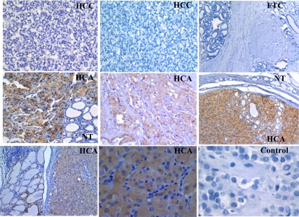
Figure 5.
Immunohistochemical analysis of KLK1 in paraffin-embedded sections of thyroid samples. HCAs exhibited strong brown immunostaining for KLK1. In contrast, FTC, most of HCC, and negative control exhibited no immunoreactivity. Adjacent normal thyroid (NT) tissue is negative for KLK1 at the same time as the tumor area is positive for PVALB.

