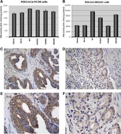
Figure 1.
PDE activity and immunohistochemistry for PDE11A. PDE activity after transfection of HEK293 cells (A) and PC3M cells (B) with wild-type (WT) and mutant PDE11A expression vectors. C, Expression of PDE11A in surrounding normal prostate cells (magnification, × 20). D, Expression of PDE11A in tumor tissue from the same PCa patient. Magnification of ×40 of PDE11A immunostaining in surrounding normal prostate cells (E) and in tumor tissue (F).

