Figure 2.

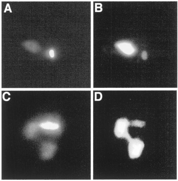
Epitope-tagged RNase H1 is present in both the nucleus and kinetoplast. (A) Wild-type C.fasciculata cells transformed with pRNH1-HA were stained with DAPI to visualize the nuclear and kinetoplast DNA. The faintly staining body is the nucleus; the brightly staining body is the kinetoplast. (B) Immunolocalization of RNH1-HA in the same cell as in (A) using a monoclonal antibody (12CA5) to the HA tag. The brightly fluorescing body is the nucleus and the faintly fluorescing body is the kinetoplast. (C) rnh1Δ strain cells transformed with pRNH1-Phleo were stained with DAPI. In this case the cell was in the process of dividing. The two faintly staining bodies are nuclei and the elongated brightly staining body is the kinetoplast. (D) Immunolocalization of RNH1-HA in the dividing cell shown in (C). Both nuclei and the kinetoplast show immunofluorescence.
