
Figure 1. GFP transgene expression in the midbrain following infusion of pseudotyped lentiviral vectors into the substantia nigra.
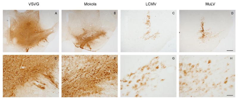
Light micrographs of coronal midbrain sections are shown, 21 days after intranigral infusion of lentiviral vectors pseudotyped with envelope proteins derived from VSV (A, E), MV (B, F), LCMV (C, G), or MuLV (D, H). GFP expression was localized immunohistochemically, with a chromogenic substrate yielding a brown product. Low magnification images (A-D; bar = 500 μm) of sections are shown for each pseudotype, in order to illustrate the topographical distribution of transgene expression. High magnification images (E-H; bar = 100 μm) of the substantia nigra show the morphology of transduced cells.