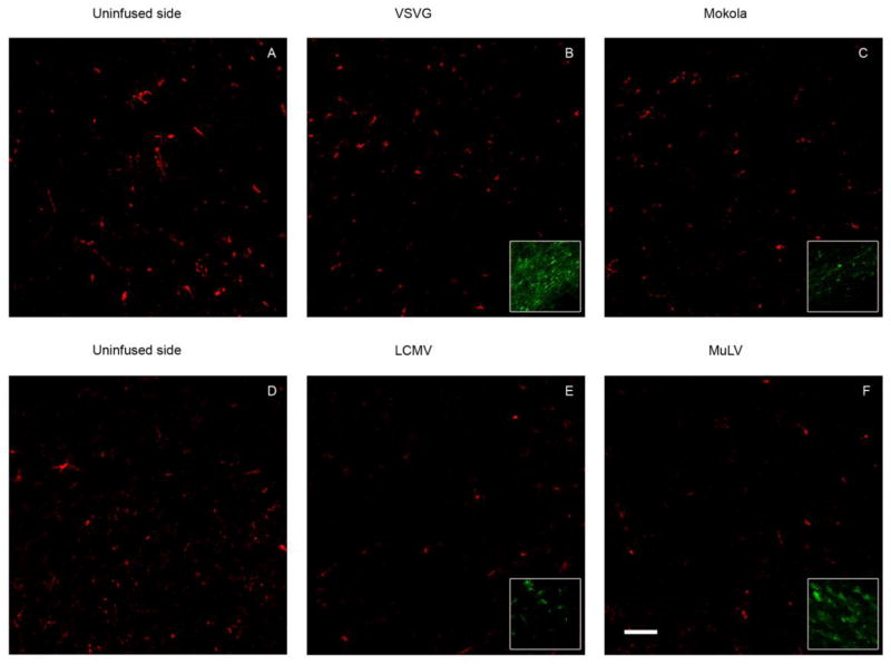
Figure 3. Pseudotyped lentiviral infusion does not induce a demonstrable immune response in the midbrain.
The images show confocal micrographs of the substantia nigra from animals that received intranigral infusions of VSV (B), MV (C), LCMV (E) or MuLV (F) pseudotyped lentiviral vectors 21 days previously (bar = 50 μm). Examples of images from the substantia nigra on the control (uninfused) side are shown for comparison (A, D). Sections were labeled by indirect immunofluorescence to detect IBA-1, a marker for microglial cells (red; A-F) and GFP in order to confirm that the IBA-1 images were obtained from regions that had been transduced by the vectors (green; inset in panels B, C, E, F). Images were acquired using identical confocal microscope settings for IBA-1 in order to allow direct comparison of the distribution and intensity of immunoreactivity. GFP images were individually adjusted in order to optimize illustration of vector transduction.

