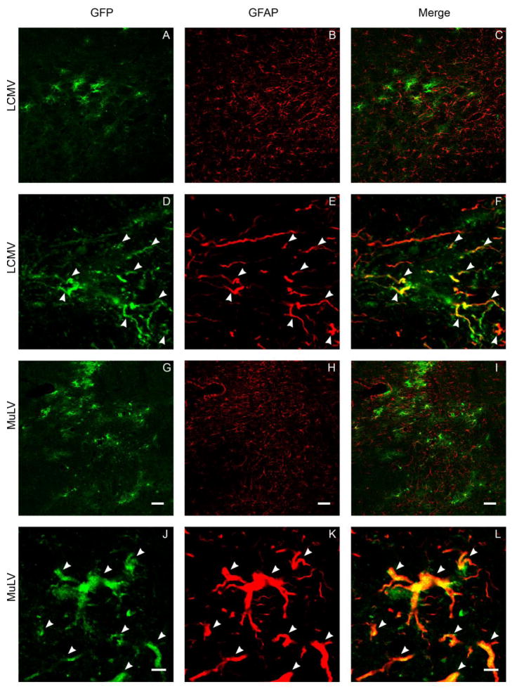
Figure 7. GFP expression in the proximal processes of astrocytes following intranigral infusion of LCMV and MuLV-pseudotyped lentiviral vectors.
The images show confocal micrographs of the substantia nigra from animals that received intranigral infusions of LCMV (A-F) or MuLV (G-L) pseudotyped lentiviral vectors 21 days previously. Sections were labeled by indirect immunofluorescence in order to detect GFP (green; A, D, G, J) and glial fibrillary acidic protein (GFAP; red; B, E, H, K). Low magnification images (A-C, G-I; bar = 50 μm) illustrate the relative topographical distributions of transduced cells and astrocytes. High magnification images (D-F, J-L bar = 5 μm) demonstrate co-localization of GFP and GFAP in the proximal processes of individual astrocytes (arrowheads).

