
Figure 9. Expression of proteins involved in cellular entry of LCMV and MuLV in the rat substantia nigra.
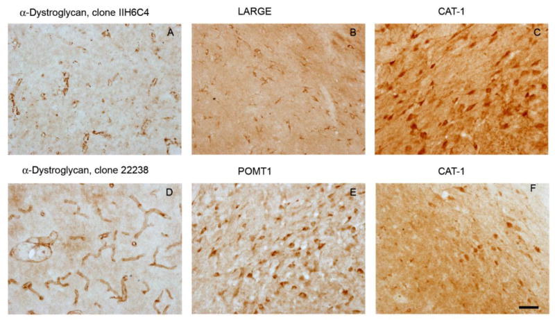
The images show light micrographs of rat substantia nigra (bar = 50 μm) labeled by immunohistochemistry with a chromogenic reaction yielding a brown product in oder to localize proteins implicated in cellular entry of LCMV (α-dystroglycan, A, D; LARGE, B; POMT1, E) or MuLV (CAT-1, C, F). Two independent antibodies for each antigen confirmed the observed expression patterns of α-dystoglycan (clones IIH6C4 A and 2238 D) and CAT-1 (Santa Cruz, C; Protein Tech, F).