
Figure 2.
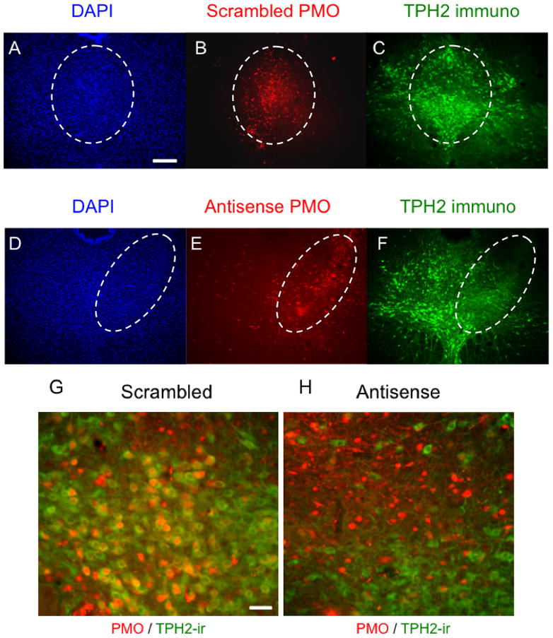
TpH immunoreactivity is reduced by PMO infusions in the midrostral DRN. Injections of scrambled PMO in the midrostral DRN (B) did not change DAPI signals (A) or TpH immunoreactivity (C). On the other hand, injections of αTpH2 PMO (E) markedly reduced TpH immunoreactivity (F) without affecting DAPI signals (D). G and H show magnified view (40X) of the scrambled and αTpH2 PMO injection site, respectively. Dashed ovals encircle the region with lissamine-PMO injection. Scale bar, 500μm (A–F), 20μm (G, H).