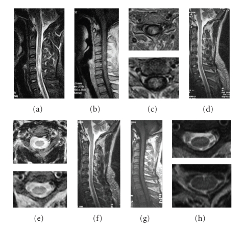
Figure 7.
A 30-year-old male suffered from left paraparesis and paresthesias for 1 month. MRI of the cervical spinal cord revealed a demyelinating lesion in T2-weighted images extending from C4 to C7. Pulse methylprednisolone and IVIG were initiated with no resolution. The MRI changes are as follows: (a) sagittal T2-weighted image reveals a high intensity signal from C4 to C7 before ATT regimen, (b) sagittal T1-weighted MRI showing light thickening of the cord with subtle intraparenchymal hyperintensity, (c) the left local lesions on axial FLAIR sequences, (d) the hyperintensity resolved following 6 months of ATT, (e) no lesion is visible on axial T2-weighted images, and (f)–(h) no lesion is visible on follow-up MRI after 2 years of treatment.

