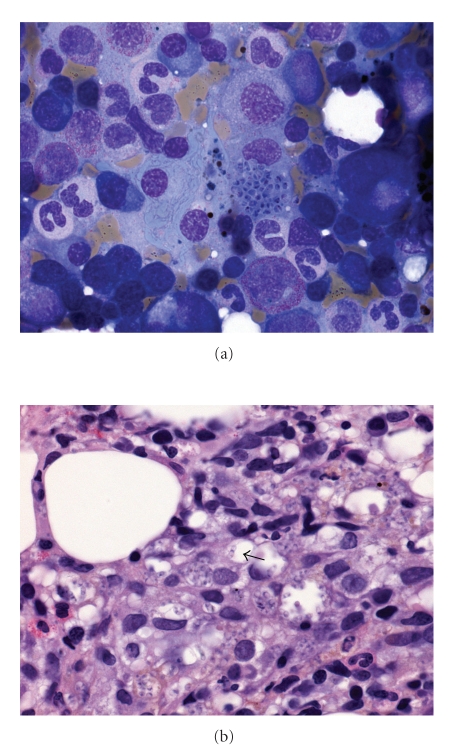
Figure 8.
(a) Marrow aspirate showing a histiocyte engorged with yeast cells with reddish pink inclusions (May Grünwald Giemsa ×1000). (b) Trephine biopsy showing histiocytic proliferation with vague granuloma formation and ingested yeast cells. Some yeast cells have transverse septum (arrow) (haematoxylin and eosin ×400).

